Sunday, February 18, 2024

Neck Pain Treatment Home Remedy

Neck pain can be a common discomfort experienced by many individuals, often resulting from poor posture, muscle strain, injury, or stress. While it can be bothersome and limit your range of motion, there are several simple and effective home remedies that can help alleviate neck pain and promote relaxation. 

These remedies aim to reduce inflammation, soothe sore muscles, and improve overall neck mobility.

Here are some potential home remedies for mild neck pain:

Rest and Relaxation:

Reduce physical activity: Take a break from strenuous activities that aggravate your neck pain.

Improve sleep hygiene: Ensure proper sleep posture with a supportive pillow to avoid stiffening.

Relaxation techniques: Practice deep breathing, meditation, or yoga to manage stress and promote healing.

Temperature Therapy:

Ice: Apply ice packs wrapped in a towel for 15-20 minutes at a time, especially in the first few days, to reduce inflammation.

Heat: After the initial inflammation subsides, use a heating pad or warm compress for 15-20 minutes to improve blood flow and relax muscles.

Stretching and Gentle Exercises:

Once the pain subsides, perform gentle neck stretches: Find guided exercises online or consult a physical therapist for safe and effective routines.

Self-massage: Use your fingertips or a tennis ball to gently massage your neck muscles and loosen tension.

Over-the-counter pain relievers:

Consider medications like ibuprofen or acetaminophen to manage pain and inflammation, but follow dosage instructions carefully.

Lifestyle adjustments:

Improve posture: Maintain good posture while sitting, standing, and sleeping. Use ergonomic furniture and a supportive pillow.

Stay hydrated: Drink plenty of water to promote circulation and muscle function.

Good Sleeping Posture:

Maintaining proper sleeping posture can prevent waking up with a stiff or sore neck. Use a supportive pillow that keeps your neck aligned with your spine, and try to avoid sleeping on your stomach, which can strain the neck muscles.

Aromatherapy:

Certain essential oils like lavender, peppermint, and eucalyptus have analgesic and anti-inflammatory properties that can help alleviate neck pain. Dilute a few drops of essential oil in a carrier oil like coconut or almond oil and massage it onto your neck for soothing relief.

Ergonomic Adjustments:

Poor posture, especially during prolonged periods of sitting or working at a computer, can contribute to neck pain. Make ergonomic adjustments to your workspace, such as ensuring your computer monitor is at eye level, using a supportive chair with proper lumbar support, and taking frequent breaks to stretch and change positions.

Remember:

These are just general suggestions, and not a substitute for professional medical advice.

Stop any home remedy that worsens your pain.

If your pain persists, worsens, or is accompanied by other symptoms, see a doctor for proper diagnosis and treatment.

Saturday, April 8, 2023

Winging of Scapula: Cause, Symptoms, Treatment, Exercise

What is a Winging of Scapula?


Winging of Scapula
Winging of Scapula

Winging of the scapula, also known as scapular winging, is a condition in which the medial or lateral border of the scapula (shoulder blade) protrudes away from the back, causing a noticeable deformity or "wing-like" appearance. This can occur as a result of weakness or paralysis of the muscles that stabilize the scapula against the rib cage, such as the serratus anterior muscle or the rhomboid muscles.

The condition can be caused by a variety of factors, including nerve damage, muscle weakness or atrophy, trauma, or underlying medical conditions such as muscular dystrophy or thoracic outlet syndrome. Symptoms of scapular winging may include pain or discomfort in the shoulder, neck or back, difficulty raising the arm or performing overhead activities, or visible protrusion of the scapula.

Treatment for scapular winging typically involves addressing the underlying cause, such as physical therapy to strengthen the affected muscles or surgery to repair nerve damage or correct skeletal abnormalities. In some cases, assistive devices such as braces or slings may also be recommended to help support the affected arm and shoulder.

Related Anatomy

The scapula, also known as the shoulder blade, is a flat, triangular bone located on the back of the thorax (chest). It is connected to the clavicle (collarbone) and the humerus (upper arm bone) by muscles and ligaments, and it articulates with the rib cage at the acromioclavicular joint.

The scapula has several bony landmarks, including the spine, acromion process, coracoid process, and glenoid cavity. The spine is a ridge of bone that runs diagonally across the posterior surface of the scapula, separating it into two unequal portions. The acromion process is a bony projection that extends from the top of the scapula and forms the tip of the shoulder. The coracoid process is a smaller bony projection that extends from the front of the scapula, just below the clavicle. The glenoid cavity is a shallow depression located on the lateral aspect of the scapula, and it forms the socket of the shoulder joint.

The muscles that attach to the scapula are responsible for stabilizing and moving the shoulder joint. These include the rotator cuff muscles (supraspinatus, infraspinatus, teres minor, and subscapularis), as well as larger muscles such as the deltoid, trapezius, serratus anterior, and rhomboids. The serratus anterior muscle, in particular, is important for stabilizing the scapula against the rib cage and preventing scapular winging. It arises from the upper eight or nine ribs and attaches to the medial border of the scapula, and it is innervated by the long thoracic nerve.

Causes of Winging of Scapula

Winging of the scapula can be caused by a variety of factors, including:

    Nerve damage: Damage to the nerves that supply the muscles responsible for stabilizing the scapula can result in scapular winging. The most common nerve involved is the long thoracic nerve, which supplies the serratus anterior muscle. Other nerves that can be affected include the spinal accessory nerve and the thoracodorsal nerve.

    Muscle weakness or atrophy: Weakness or atrophy of the muscles that stabilize the scapula against the rib cage, such as the serratus anterior or the rhomboid muscles, can also cause scapular winging. This can be due to a variety of factors, including disuse, nerve damage, or muscle disorders such as muscular dystrophy.

    Trauma: Direct trauma to the scapula or the surrounding area can cause damage to the muscles or nerves that control scapular movement, resulting in scapular winging.

    Underlying medical conditions: Certain medical conditions such as thoracic outlet syndrome, myopathies, or neuropathies can lead to scapular winging.

    Surgery: Surgery involving the shoulder or the chest wall can sometimes result in scapular winging as a complication.

It's important to identify the underlying cause of scapular winging in order to determine the most appropriate treatment approach.

Symptoms of Winging of Scapula

Symptoms of winging of the scapula can vary depending on the underlying cause, but may include:

    Visible protrusion of the scapula: The medial or lateral border of the scapula may protrude away from the back, giving a noticeable deformity or "wing-like" appearance.

    Pain or discomfort: Patients may experience pain or discomfort in the shoulder, neck, or upper back, particularly when using the affected arm or performing overhead activities.

    Weakness: Weakness or difficulty with arm elevation or pushing tasks may be present, as the muscles responsible for stabilizing the scapula may be compromised.

    Limited range of motion: Patients may have limited range of motion in the shoulder joint, particularly when trying to lift the arm above shoulder height or behind the back.

    Muscle atrophy: Over time, the muscles responsible for stabilizing the scapula may become smaller or weaker, resulting in muscle atrophy.

It's important to seek medical attention if you experience any of these symptoms, as early diagnosis and treatment can help prevent complications and improve outcomes.

Differential Diagnosis

Winging of the scapula is a relatively rare condition, and there are several conditions that may present with similar symptoms. Differential diagnosis for scapular winging may include:

    Rotator cuff injury: Injury or tears to the rotator cuff muscles can cause pain, weakness, and limited range of motion in the shoulder, which may mimic the symptoms of scapular winging.

    Shoulder impingement syndrome: This condition occurs when the tendons or bursae in the shoulder become compressed or irritated, resulting in pain and limited range of motion. Shoulder impingement can also cause weakness in the affected arm, which may be mistaken for scapular winging.

    Thoracic outlet syndrome: This condition occurs when the nerves or blood vessels that pass through the thoracic outlet become compressed or pinched, resulting in pain, weakness, and numbness in the arm and hand. In some cases, thoracic outlet syndrome may also cause scapular winging.

    Cervical radiculopathy: This condition occurs when a nerve root in the cervical spine becomes compressed or irritated, resulting in pain, weakness, and numbness in the shoulder, arm, and hand. Cervical radiculopathy can sometimes be mistaken for scapular winging.

    Muscular dystrophy: Muscular dystrophy is a group of inherited disorders that cause progressive muscle weakness and wasting, which can result in scapular winging in some cases.

    Trauma or injury: Trauma or injury to the shoulder or back can cause pain, weakness, and limited range of motion, which may mimic the symptoms of scapular winging.

It's important to consult with a healthcare professional if you are experiencing any symptoms of scapular winging or if you have any concerns about your shoulder or back health. A proper diagnosis is crucial for effective treatment and management of your symptoms.

Diagnosis

The diagnosis of scapular winging typically involves a thorough physical examination and a review of the patient's medical history. The healthcare provider may ask the patient about the onset and duration of symptoms, any history of injury or trauma, and any underlying medical conditions that may contribute to the symptoms.

During the physical examination, the healthcare provider will assess the patient's range of motion, muscle strength, and the appearance of the scapula. They may ask the patient to perform specific movements or exercises to evaluate the function of the muscles responsible for scapular stability. The healthcare provider may also palpate the scapula and surrounding muscles to identify any areas of tenderness or weakness.

Diagnostic imaging, such as X-rays, MRI, or CT scans, may also be ordered to evaluate the bones, joints, and soft tissues in the affected area. Electromyography (EMG) may also be used to assess nerve and muscle function.

If scapular winging is suspected, the healthcare provider may perform additional tests to identify the underlying cause. This may include blood tests, nerve conduction studies, or a biopsy of the affected muscles.

The diagnosis of scapular winging can be complex, and it's important to consult with a healthcare provider who specializes in the evaluation and treatment of shoulder and back conditions. A proper diagnosis is crucial for developing an effective treatment plan and improving outcomes.

Treatment of Winging of Scapula

The treatment of scapular winging depends on the underlying cause and the severity of the symptoms. In some cases, conservative management may be sufficient, while in other cases, surgery may be necessary. Some common treatment options for scapular winging include:

    Physical therapy: Physical therapy can help improve range of motion, strengthen the muscles responsible for scapular stability, and improve overall shoulder function. Your physical therapist may recommend specific exercises and stretches to target the affected muscles and improve your symptoms.

    Bracing: A scapular brace can help support the shoulder and improve scapular stability. Depending on the severity of the scapular winging, the healthcare provider may recommend an off-the-shelf or custom-made brace.

    Medications: Nonsteroidal anti-inflammatory drugs (NSAIDs) may be recommended to help manage pain and inflammation associated with scapular winging.

    Injections: Corticosteroid injections may be recommended to reduce inflammation and pain in the affected area.

    Surgery: In severe cases of scapular winging, surgery may be necessary to correct the underlying cause of the condition. The surgical approach will depend on the cause of the scapular winging, and may involve repositioning or reattaching the affected muscles, removing any abnormalities in the bone, or repairing any damage to the nerves or blood vessels in the area.

It's important to consult with a healthcare provider to determine the most appropriate treatment plan for your specific case of scapular winging. With proper treatment and management, most patients with scapular winging can experience significant improvement in their symptoms and function.

Physiotherapy Treatment in Winging of Scapula

Physical therapy is an important component of the treatment plan for scapular winging. A physical therapist can help improve scapular stability and shoulder function through specific exercises and manual techniques. The goals of physical therapy for scapular winging may include:

    Improving scapular stability: The physical therapist may recommend exercises to strengthen the muscles responsible for scapular stability, such as the serratus anterior, trapezius, and rhomboids. Strengthening these muscles can help improve the position and function of the scapula, which can improve shoulder function and reduce symptoms.

    Improving range of motion: The physical therapist may recommend stretches and exercises to improve range of motion in the shoulder and upper back. This can help improve overall shoulder function and reduce pain and stiffness.

    Correcting postural abnormalities: The physical therapist may assess the patient's posture and recommend exercises and techniques to correct any postural abnormalities that may be contributing to the scapular winging.

    Pain management: The physical therapist may use manual techniques, such as massage or mobilization, to help reduce pain and improve tissue mobility in the affected area.

    Education: The physical therapist can educate the patient on proper body mechanics and techniques for performing daily activities to help prevent further injury or aggravation of the condition.

The specific treatment plan will depend on the underlying cause of the scapular winging and the severity of the symptoms. The physical therapist will work with the patient to develop an individualized treatment plan based on their specific needs and goals.

Exercise for Winging of Scapula

Exercises that focus on strengthening the serratus anterior, trapezius, and rhomboid muscles can be beneficial for improving scapular stability and reducing symptoms of scapular winging. Here are some exercises that may be recommended by a physical therapist or healthcare provider:

    Wall slides: Stand with your back against a wall, with your feet shoulder-width apart. Place your hands on the wall at shoulder height, with your elbows bent to 90 degrees. Slowly slide your arms up the wall, keeping your elbows and wrists in contact with the wall. Hold for a few seconds, then slowly lower your arms back down. Repeat for 10-15 repetitions.

    Scapular push-ups: Start in a plank position, with your arms straight and your shoulders directly over your wrists. Slowly lower your body towards the ground, while simultaneously squeezing your shoulder blades together. Push back up to the starting position, while maintaining the squeeze in your shoulder blades. Repeat for 10-15 repetitions.

    Prone Y-T-W-L: Lie face down on a mat or bench, with your arms hanging down towards the floor. Lift your arms up and out to the side to form a Y shape, then lower back down. Lift your arms up and out to the side to form a T shape, then lower back down. Lift your arms up and out to the side to form a W shape, then lower back down. Finally, lift your arms up and out to the side to form an L shape, then lower back down. Repeat for 10-15 repetitions of each position.

    Resistance band rows: Attach a resistance band to a stable object, such as a door or pole. Stand facing the object, with the resistance band in both hands. Bend your knees slightly and lean forward from the hips, keeping your back straight. Pull the resistance band towards your chest, squeezing your shoulder blades together. Slowly release back to the starting position. Repeat for 10-15 repetitions.

It's important to work with a healthcare provider or physical therapist to determine the most appropriate exercises for your specific case of scapular winging. They can help develop an individualized exercise plan based on your specific needs and goals.

Complications

In some cases, untreated or improperly managed scapular winging can lead to complications, such as:

    Shoulder instability: Scapular winging can lead to instability of the shoulder joint, which can increase the risk of dislocation or subluxation (partial dislocation) of the shoulder.

    Rotator cuff injuries: Scapular winging can also increase the risk of rotator cuff injuries, as the muscles that stabilize the scapula and the rotator cuff muscles work together to support shoulder movement.

    Chronic pain: Scapular winging can cause chronic pain in the shoulder, neck, and upper back, which can affect daily activities and quality of life.

    Decreased range of motion: If scapular winging is left untreated, it can lead to a decreased range of motion in the shoulder joint, which can limit the ability to perform daily activities and participate in sports or other physical activities.

    Muscle atrophy: Chronic scapular winging can also lead to muscle atrophy (wasting) in the affected muscles, which can further weaken the shoulder and limit function.

It's important to seek prompt medical attention if you experience symptoms of scapular winging, such as shoulder pain, weakness, or a protruding shoulder blade. Early diagnosis and proper management can help prevent complications and improve outcomes.

How to Prevent Winging of Scapula?

Scapular winging can be prevented or minimized by:

    Maintaining good posture: Maintaining good posture can help prevent scapular winging by keeping the shoulder blades in the correct position and reducing strain on the muscles that support the scapula.

    Strengthening the shoulder and back muscles: Strengthening the muscles that support the shoulder and back can help improve scapular stability and prevent winging.

    Avoiding overuse or repetitive motions: Overuse or repetitive motions of the shoulder and upper back can increase the risk of scapular winging. Taking breaks, modifying activities, or using proper technique can help prevent injury and reduce the risk of winging.

    Addressing underlying medical conditions: Certain medical conditions, such as nerve injuries or muscular dystrophy, can increase the risk of scapular winging. Treating these conditions can help prevent or minimize winging.

    Seeking prompt medical attention: If you experience shoulder pain, weakness, or other symptoms of scapular winging, seek prompt medical attention. Early diagnosis and management can help prevent complications and improve outcomes.

It's important to work with a healthcare provider or physical therapist to develop an individualized prevention plan based on your specific needs and risk factors.

What are the 2 types of scapular winging?

The two types of scapular winging are:

    Medial scapular winging: In medial scapular winging, the inner border of the scapula (the part closest to the spine) lifts away from the ribcage. This is typically caused by damage to the long thoracic nerve, which innervates the serratus anterior muscle.

    Lateral scapular winging: In lateral scapular winging, the outer border of the scapula (the part furthest from the spine) lifts away from the ribcage. This is typically caused by damage to the accessory nerve or the trapezius muscle.

How do you test for scapular winging?

Scapular winging can be tested using the following methods:
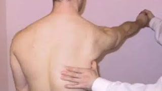

Winging of Scapula test

Wall push-up test: The patient is asked to stand with their hands on a wall and perform a push-up motion while the examiner observes the movement of the scapula. If the scapula lifts away from the ribcage during the movement, this may indicate scapular winging.

    Scapular assist test: The patient is asked to hold a weight or resistance band with both hands and lift it overhead while the examiner observes the movement of the scapula. If the scapula lifts away from the ribcage during the movement, this may indicate scapular winging.

    Scapular retraction test: The patient is asked to hold their arms out in front of them and retract their shoulder blades while the examiner observes the movement of the scapula. If the scapula lifts away from the ribcage during the movement, this may indicate scapular winging.

    Manual muscle testing: The strength of the muscles that support the scapula, such as the serratus anterior and trapezius muscles, can be tested using manual muscle testing. Weakness or muscle atrophy may indicate scapular winging.

If scapular winging is suspected based on these tests, imaging studies such as X-rays, MRI, or electromyography (EMG) may be ordered to confirm the diagnosis and determine the underlying cause.

Can winged scapula be fixed by exercise?

In some cases, scapular winging can be improved or even corrected through exercise. The effectiveness of exercise in treating scapular winging depends on the underlying cause and severity of the condition.

For example, if scapular winging is caused by weakness or imbalances in the muscles that support the scapula, targeted exercises can help improve muscle strength and function. Strengthening exercises may include exercises for the serratus anterior, such as wall push-ups, shoulder protraction exercises, and scapular push-ups, as well as exercises for the trapezius and rhomboid muscles.

However, if scapular winging is caused by nerve damage or structural abnormalities, exercise alone may not be sufficient to correct the problem. In these cases, other interventions such as surgery or physical therapy may be necessary.

It's important to work with a healthcare provider or physical therapist to develop an individualized exercise program based on your specific needs and the underlying cause of your scapular winging. They can also monitor your progress and adjust your exercise program as needed to ensure that you are making progress and not causing further damage.

How long is recovery for winged scapula?

The recovery time for scapular winging depends on the underlying cause and severity of the condition, as well as the treatment approach taken.

If scapular winging is caused by a nerve injury, recovery time may be longer and may involve a combination of surgery, physical therapy, and medication. Recovery time may range from several weeks to several months or longer, depending on the severity of the nerve damage and the individual's response to treatment.

If scapular winging is caused by muscle weakness or imbalances, recovery time may be shorter and may involve targeted exercises to strengthen the muscles that support the scapula. The length of recovery time will depend on the severity of the muscle weakness and how well the individual responds to exercise therapy.

It's important to work closely with a healthcare provider or physical therapist to develop a treatment plan that is tailored to your individual needs and to monitor your progress throughout the recovery process. They can also provide guidance on how to prevent future episodes of scapular winging and how to safely return to your normal activities.

What is the fastest way to fix a winged scapula?

There is no single "fastest" way to fix a winged scapula as the treatment approach will depend on the underlying cause and severity of the condition. However, there are some steps that can be taken to address the issue as quickly and effectively as possible:

    Consult with a healthcare provider: If you suspect that you have scapular winging, it's important to consult with a healthcare provider who can perform a thorough evaluation and determine the underlying cause of the condition. Based on the diagnosis, the provider can recommend an appropriate treatment plan.

    Begin physical therapy: Physical therapy can be an effective way to treat scapular winging caused by muscle weakness or imbalances. A physical therapist can develop an individualized exercise program to target the affected muscles and help improve strength and function. Starting physical therapy as soon as possible can help speed up the recovery process.

    Consider surgery: In some cases, scapular winging may be caused by nerve damage or structural abnormalities that require surgical intervention. If this is the case, your healthcare provider may recommend surgery as the fastest way to address the issue.

    Address contributing factors: In addition to exercise and/or surgery, it's important to address any contributing factors that may be exacerbating the condition, such as poor posture or improper lifting techniques. This can help prevent future episodes of scapular winging and promote faster healing.

It's important to remember that there is no one-size-fits-all approach to treating scapular winging, and recovery time can vary widely depending on the individual and the underlying cause of the condition. Working closely with a healthcare provider and physical therapist can help ensure the best possible outcome.

What nerve damage causes winging scapula?

The nerve damage that most commonly causes winging scapula is damage to the long thoracic nerve. This nerve innervates the serratus anterior muscle, which helps to stabilize the scapula against the ribcage during shoulder movements. If the long thoracic nerve is damaged, the serratus anterior muscle may become weak or paralyzed, leading to scapular winging.

The long thoracic nerve can be damaged by a number of factors, including trauma or injury to the shoulder or neck, surgical procedures in the area, or repetitive overhead activities such as throwing or serving. In some cases, the cause of the nerve damage may be unknown.

It's important to note that scapular winging can also be caused by other factors such as muscle weakness or imbalances, structural abnormalities, or neurological disorders. Therefore, a thorough evaluation by a healthcare provider is necessary to determine the underlying cause of the condition and develop an appropriate treatment plan.

What doctor treats winged scapula?

Several healthcare providers may be involved in the diagnosis and treatment of winged scapula, depending on the underlying cause of the condition. Some healthcare providers who may be involved in the care of patients with winged scapula include:

    Orthopedic surgeon: An orthopedic surgeon is a medical doctor who specializes in the diagnosis and treatment of musculoskeletal conditions, including those affecting the shoulder and scapula. They may be involved in the surgical management of winged scapula, especially if the condition is caused by nerve damage or structural abnormalities.

    Neurologist: A neurologist is a medical doctor who specializes in the diagnosis and treatment of conditions affecting the nervous system, including nerve damage that can cause winged scapula.

    Physical therapist: A physical therapist is a healthcare provider who specializes in the evaluation and treatment of musculoskeletal conditions using exercise therapy, manual therapy, and other interventions. They may develop an exercise program to help strengthen the muscles supporting the scapula and improve function.

    Sports medicine physician: A sports medicine physician is a medical doctor who specializes in the diagnosis and treatment of sports-related injuries and conditions. They may be involved in the care of athletes or individuals who develop winged scapula as a result of repetitive overhead activities or other athletic endeavors.

    Primary care physician: A primary care physician is a medical doctor who provides general medical care and may be involved in the initial evaluation and management of winged scapula.

It's important to work closely with a healthcare provider or team of providers to determine the underlying cause of winged scapula and develop an appropriate treatment plan.

Summary

Winged scapula is a condition where the shoulder blade sticks out from the back, which can cause pain, weakness, and limited mobility. It can be caused by various factors, including nerve damage, muscle weakness or imbalances, structural abnormalities, or neurological disorders. The long thoracic nerve is the most common nerve that causes winged scapula due to damage or injury. The diagnosis of winged scapula involves a physical exam, medical history, and imaging tests, and treatment may include physical therapy, exercises, medication, or surgery. Several healthcare providers may be involved in the diagnosis and treatment of winged scapula, including an orthopedic surgeon, neurologist, physical therapist, sports medicine physician, and primary care physician. Preventive measures for winged scapula include good posture, proper form during exercise or sports, and avoiding repetitive overhead activities.

Tuesday, March 28, 2023

Deltoid muscle anatomy Origin, Insertion, Function, Exercise


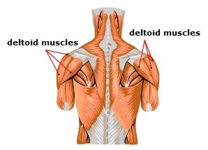
Deltoid Muscle
Deltoid Muscle

The deltoid muscle is a large, triangular muscle located in the shoulder region. It is named for its shape, which resembles a delta or the Greek letter delta (Δ). The deltoid muscle is composed of three distinct regions or heads: the anterior (front), middle, and posterior (back) heads.

The anterior head originates from the lateral third of the clavicle, while the middle head originates from the acromion process of the scapula. The posterior head originates from the spine of the scapula. All three heads converge to form a single tendon, which inserts into the deltoid tuberosity of the humerus bone.

The deltoid muscle is innervated by the axillary nerve, which arises from the brachial plexus. It is responsible for several movements of the shoulder joint, including abduction (lifting the arm away from the body), flexion (raising the arm in front of the body), and extension (moving the arm backward). Additionally, it assists in medial and lateral rotation of the arm.

Overall, the deltoid muscle is an important muscle for the stability and mobility of the shoulder joint.

Origin of Deltoid muscle

The deltoid muscle has three distinct regions or heads that have different origins:

  1. The anterior (front) head originates from the lateral third of the clavicle.
  2. The middle head originates from the acromion process of the scapula and the lateral border of the spine of the scapula.
  3. The posterior (back) head originates from the inferior edge of the crest of the spine of the scapula.

All three heads of the deltoid converge to form a single tendon, which inserts into the deltoid tuberosity of the humerus bone. The deltoid muscle is innervated by the axillary nerve, which arises from the brachial plexus.

Insertion

The deltoid muscle inserts into the deltoid tuberosity of the humerus bone, which is located on the lateral side of the shaft of the humerus, about one-third of the distance from the proximal end (shoulder end) to the distal end (elbow end) of the bone.

The deltoid muscle's tendon is broad and flat, and it covers the superior and lateral aspects of the humeral head. It is important for the stability and mobility of the shoulder joint, and it helps to hold the humeral head in the glenoid fossa of the scapula during shoulder movement.

In addition to the deltoid tuberosity of the humerus, the deltoid muscle also has some weak insertions into the capsule of the shoulder joint, the acromion process of the scapula, and the spine of the scapula. These additional insertions may provide some extra stability to the shoulder joint.

Nerve supply

The deltoid muscle is innervated by the axillary nerve, which arises from the posterior cord of the brachial plexus. The axillary nerve arises from the spinal nerves C5 and C6, and it passes through the quadrilateral space, a small opening in the posterior aspect of the shoulder, to reach the deltoid muscle.

The axillary nerve supplies both motor and sensory innervation to the deltoid muscle. It provides the motor innervation that allows the deltoid muscle to contract and produce movement at the shoulder joint, including abduction (lifting the arm away from the body), flexion (raising the arm in front of the body), and extension (moving the arm backward). The axillary nerve also provides sensory innervation to the skin overlying the deltoid muscle, as well as the joint capsule of the shoulder joint.

Injury or damage to the axillary nerve can result in weakness or paralysis of the deltoid muscle, which can lead to difficulty with shoulder movement and stability.

Blood supply

The deltoid muscle receives its blood supply from several arteries, including the posterior circumflex humeral artery, the deltoid branch of the thoracoacromial artery, and the suprascapular artery.

The posterior circumflex humeral artery is the main arterial supply to the deltoid muscle, and it arises from the third part of the axillary artery. It runs around the surgical neck of the humerus, along with the axillary nerve, and then divides into several branches that supply the deltoid muscle.

The deltoid branch of the thoracoacromial artery is a small branch that arises from the thoracoacromial artery, which is a branch of the axillary artery. It supplies the anterior part of the deltoid muscle.

The suprascapular artery is another small branch of the subclavian artery that supplies the shoulder joint and the muscles around it, including the deltoid muscle.

These arteries provide a rich blood supply to the deltoid muscle, which is important for the muscle's function and recovery after injury.

Function of Deltoid muscle

The deltoid muscle is responsible for several movements of the shoulder joint and provides stability to the joint during arm movements. Its functions include:

  • Shoulder abduction: The deltoid muscle is the primary muscle that lifts the arm away from the body in the plane of the scapula. When the deltoid contracts, it pulls the humerus bone upward and away from the body, allowing the arm to move outward and upward.
  • Shoulder flexion: The anterior fibers of the deltoid muscle assist in flexing the arm at the shoulder joint, allowing the arm to move forward and upward in front of the body.
  • Shoulder extension: The posterior fibers of the deltoid muscle assist in extending the arm at the shoulder joint, allowing the arm to move backward behind the body.
  • Shoulder horizontal abduction: The middle fibers of the deltoid muscle are responsible for moving the arm outward away from the midline of the body.
  • Shoulder horizontal adduction: The anterior fibers of the deltoid muscle are involved in moving the arm inward towards the midline of the body.
  • Medial and lateral rotation: The deltoid muscle also assists in rotating the arm medially (inward) and laterally (outward).

Overall, the deltoid muscle is a critical muscle for shoulder joint mobility and stability, and it is essential for many activities that involve arm movement, such as throwing, reaching, lifting, and pushing.

Clinical significance

The deltoid muscle can be affected by various injuries and conditions that can result in pain, weakness, or limited range of motion. Some of the clinical significance of the deltoid muscle include:

  • Rotator cuff injuries: The rotator cuff muscles and tendons, which surround the shoulder joint, are often injured in sports or from overuse. These injuries can result in pain, weakness, and limited mobility of the shoulder joint, which can affect the function of the deltoid muscle.
  • Shoulder impingement syndrome: This condition occurs when the tendons of the rotator cuff and the bursa (a fluid-filled sac) become compressed between the bones of the shoulder joint, causing pain, inflammation, and reduced mobility. Shoulder impingement syndrome can affect the function of the deltoid muscle and limit its ability to lift the arm.
  • Shoulder dislocation: When the humerus bone is forced out of the shoulder joint, it can damage the deltoid muscle, as well as other muscles and tissues around the shoulder. Shoulder dislocations can cause pain, swelling, weakness, and limited range of motion in the shoulder joint.
  • Nerve injuries: The axillary nerve, which innervates the deltoid muscle, can be injured or compressed due to trauma or overuse, resulting in weakness or paralysis of the deltoid muscle.
  • Muscle strains or tears: The deltoid muscle can be strained or torn due to overuse or trauma, resulting in pain, swelling, weakness, and limited range of motion of the shoulder joint.

In addition to these conditions, the deltoid muscle can also be affected by diseases such as muscular dystrophy or amyotrophic lateral sclerosis (ALS), which can result in muscle weakness and atrophy (wasting). Overall, understanding the anatomy and function of the deltoid muscle is important for the diagnosis and treatment of various shoulder-related injuries and conditions.

Stretching exercises of Deltoid muscle

Stretching the deltoid muscle can help improve shoulder mobility, reduce pain, and prevent injuries. Here are a few examples of stretching exercises for the deltoid muscle:

  • Standing wall stretch: Stand facing a wall with your feet hip-width apart. Place one hand on the wall, with your arm straight and your fingers pointing upward. Slowly turn your body away from the wall, keeping your arm straight, until you feel a stretch in your shoulder. Hold for 15-30 seconds, then repeat on the other side.
  • Cross-body stretch: Stand or sit with your back straight. Bring one arm across your chest and hold it with the opposite hand. Gently pull the arm toward your body until you feel a stretch in your shoulder. Hold for 15-30 seconds, then repeat on the other side.
  • Doorway stretch: Stand in a doorway with your arms at your sides. Place your hands on the sides of the doorway at shoulder height. Step forward with one foot, keeping your arms straight and your hands on the doorway. Lean forward until you feel a stretch in your shoulders. Hold for 15-30 seconds, then step back and repeat on the other side.
  • Shoulder circles: Stand with your feet hip-width apart and your arms at your sides. Slowly raise your shoulders up toward your ears, then roll them back and down in a circular motion. Repeat for 10-15 repetitions, then reverse the direction.

It's important to remember to stretch gently and gradually, without bouncing or jerking. If you have any shoulder pain or discomfort, consult a healthcare professional before starting any stretching or exercise program.

Strengthening exercises for Deltoid muscle

Strengthening the deltoid muscle can help improve shoulder stability and function, increase mobility, and prevent injuries. Here are a few examples of strengthening exercises for the deltoid muscle:

  • Shoulder press: Stand or sit with your back straight and your feet hip-width apart. Hold a dumbbell or a barbell in each hand at shoulder height, with your palms facing forward. Raise the weights straight up above your head, then lower them back down to shoulder height. Repeat for 8-12 repetitions.
  • Lateral raise: Stand or sit with your back straight and your feet hip-width apart. Hold a dumbbell or a weight plate in each hand at your sides, with your palms facing inward. Raise your arms straight out to the sides until they are parallel to the floor, then lower them back down to your sides. Repeat for 8-12 repetitions.
  • Bent-over raise: Stand with your feet hip-width apart and your knees slightly bent. Hold a dumbbell in each hand, with your palms facing inward. Bend forward at the waist, keeping your back straight and your arms hanging down in front of you. Raise your arms straight out to the sides until they are parallel to the floor, then lower them back down. Repeat for 8-12 repetitions.
  • Upright row: Stand with your feet hip-width apart and hold a barbell or a pair of dumbbells in front of your thighs, with your palms facing your body. Lift the weights straight up to your chin, keeping your elbows out to the sides. Lower the weights back down to your thighs. Repeat for 8-12 repetitions.

It's important to use proper form and technique when performing these exercises, and to start with a weight that is appropriate for your strength level. Gradually increase the weight and the number of repetitions as you get stronger. If you have any shoulder pain or discomfort, consult a healthcare professional before starting any strengthening or exercise program.

Conclusion

The deltoid muscle is a large, triangular muscle located in the shoulder region. It consists of three parts: the anterior, middle, and posterior deltoids, which have different origins, insertions, and functions. The deltoid muscle is responsible for a wide range of shoulder movements, including flexion, extension, abduction, adduction, and rotation.

Stretching and strengthening exercises for the deltoid muscle can help improve shoulder mobility, stability, and function, and prevent injuries. Examples of stretching exercises include standing wall stretch, cross-body stretch, doorway stretch, and shoulder circles. 

Examples of strengthening exercises include shoulder press, lateral raise, bent-over raise, and upright row. It's important to use proper form and technique when performing these exercises, and to start with a weight that is appropriate for your strength level. If you have any shoulder pain or discomfort, consult a healthcare professional before starting any stretching or exercise program.

Monday, March 20, 2023

Neck Pain Overview

What is Neck pain?


Neck Pain
Neck Pain

Neck pain is a common condition characterized by discomfort or pain in the neck region. It can be caused by a variety of factors, including poor posture, muscle strain or injury, arthritis, disc herniation, spinal stenosis, or nerve impingement. 

The pain can range from mild to severe and may be accompanied by other symptoms such as stiffness, numbness or tingling, headaches, or shoulder pain. Neck pain can be acute, meaning it comes on suddenly and lasts for a short period of time, or it can be chronic, lasting for weeks or even months. Treatment options may include physical therapy, pain medication, rest, and in some cases, surgery.

What are the causes of Neck pain?

There are many possible causes of neck pain. Some of the most common causes include:

Poor posture: Maintaining an incorrect posture while sitting or standing can put undue pressure on the neck muscles and cause pain.

Muscle strain or injury: Overuse of the neck muscles or sudden twisting or jerking of the neck can cause strain or injury to the muscles or ligaments in the neck.

Arthritis: Arthritis can cause inflammation in the joints of the neck, leading to pain and stiffness.

Disc herniation: When one of the discs between the vertebrae in the neck ruptures or bulges out, it can put pressure on nearby nerves and cause pain.

Spinal stenosis: A narrowing of the spinal canal in the neck can cause pressure on the spinal cord and nerve roots, leading to pain.

Nerve impingement: When a nerve in the neck becomes pinched or compressed, it can cause pain, numbness, or tingling in the neck and arms.

Trauma or injury: A sudden impact, such as a car accident or fall, can cause neck injury and pain.

Medical conditions: Certain medical conditions such as fibromyalgia, meningitis, or cancer can also cause neck pain.

What are The symptoms of Neck pain?

The symptoms of neck pain can vary depending on the cause and severity of the pain, but may include:

Pain or stiffness in the neck, shoulders, or upper back

Headaches, especially at the base of the skull

Limited range of motion or difficulty moving the neck

Muscle spasms in the neck and upper back

Numbness, tingling, or weakness in the arms, hands, or fingers

Dizziness or lightheadedness

Difficulty swallowing or breathing

Tenderness or swelling in the neck

Grinding or popping sounds when moving the neck

Pain that worsens with movement or certain activities.

It is important to note that neck pain can sometimes be a sign of a more serious medical condition, such as a herniated disc, spinal cord injury, or infection, and may require urgent medical attention.

Difference Diagnosis

Neck pain can be caused by a variety of factors, and the diagnosis can vary based on the underlying cause. Some common differential diagnoses for neck pain include:

Cervical strain: This is a common cause of neck pain that occurs due to muscle strain or overuse.

Cervical radiculopathy: This condition occurs when a nerve in the neck becomes pinched or compressed, causing pain, numbness, or tingling in the neck and arms.

Cervical herniated disc: This condition occurs when the soft, jelly-like material inside a disc in the neck ruptures or bulges out, putting pressure on nearby nerves and causing pain.

Cervical osteoarthritis: This condition occurs when the joints in the neck become inflamed, leading to pain, stiffness, and reduced range of motion.

Cervical spinal stenosis: This condition occurs when the spinal canal in the neck narrows, putting pressure on the spinal cord and nerves and causing pain, weakness, and numbness.

Tension headache: This type of headache can cause pain and pressure in the neck and head, often due to muscle tension or stress.

Meningitis: This is a serious condition in which the membranes surrounding the brain and spinal cord become inflamed, causing neck pain, fever, and other symptoms.

Diagnosis of neck pain often involves a physical exam, medical history, imaging tests such as X-rays or MRI, and possibly other diagnostic tests such as nerve conduction studies. The specific diagnosis will depend on the underlying cause of the neck pain.

Treatment of Neck pain

The treatment for neck pain depends on the underlying cause and severity of the pain. Some common treatment options include:

Pain medication: Over-the-counter pain medications such as acetaminophen or nonsteroidal anti-inflammatory drugs (NSAIDs) can help relieve mild to moderate neck pain.

Physical therapy Treatment: Exercises and stretches designed to improve neck mobility and strengthen the muscles can help reduce pain and prevent future injury.

Heat or ice therapy: Applying heat or ice to the affected area can help reduce pain and inflammation.

Massage therapy: Gentle massage can help relieve muscle tension and reduce pain.

Chiropractic manipulation: This involves the use of manual manipulation techniques to improve neck mobility and reduce pain.

Acupuncture: This is a technique that involves the insertion of thin needles into specific points on the body to help reduce pain and inflammation.

Surgery: In some cases, surgery may be necessary to treat underlying conditions such as herniated discs or spinal stenosis.

It is important to consult with a healthcare professional to determine the best course of treatment for your specific condition. In addition to treatment, lifestyle changes such as maintaining good posture, getting regular exercise, and reducing stress can help prevent future neck pain.

How to Prevent Neck pain?

Here are some tips to help prevent neck pain:

Practice good posture: Maintaining good posture can help reduce strain on the neck muscles and prevent neck pain.

Take frequent breaks: If you spend a lot of time sitting at a desk or using a computer, take frequent breaks to stand up, stretch, and move around.

Use ergonomic equipment: Ensure that your workspace is set up in a way that promotes good posture and reduces strain on the neck.

Exercise regularly: Regular exercise can help improve neck strength and flexibility, which can help prevent injury and reduce pain.

Use proper lifting techniques: When lifting heavy objects, use proper techniques such as bending at the knees and keeping the object close to your body to avoid straining the neck muscles.

Manage stress: Stress can cause muscle tension, which can lead to neck pain. Practice stress-reduction techniques such as meditation, deep breathing, or yoga.

Sleep on a supportive mattress and pillow: A supportive mattress and pillow can help maintain proper spinal alignment and prevent neck pain.

By following these tips, you can reduce your risk of developing neck pain and maintain good neck health.

Exercises for Neck pain

Here are some exercises that can help relieve and prevent neck pain:

Neck stretches: Tilt your head to the left and hold for 10 seconds, then tilt to the right and hold for 10 seconds. Repeat 5-10 times. Then, tilt your head forward and hold for 10 seconds, then tilt your head backward and hold for 10 seconds. Repeat 5-10 times.

Shoulder rolls: Roll your shoulders forward 10 times, then roll them backward 10 times.

Chin tucks: While sitting or standing, pull your chin straight back toward your neck and hold for 5 seconds. Repeat 10 times.

Side bends: Tilt your head to the left and place your left hand on top of your head. Gently pull your head toward your left shoulder and hold for 10 seconds. Repeat on the right side.

Resistance exercises: Place your hand against your forehead and gently push your head forward, using your neck muscles to resist the pressure. Hold for 5 seconds, then release. Repeat 10 times. Then, place your hand on the back of your head and gently push your head backward, again resisting the pressure with your neck muscles. Hold for 5 seconds, then release. Repeat 10 times.

Shoulder blade squeezes: Sit or stand with your arms at your sides. Squeeze your shoulder blades together and hold for 5 seconds, then release. Repeat 10 times.

Remember to consult with a healthcare professional before starting any exercise program, especially if you have a history of neck pain or injury.

Wednesday, June 19, 2019

Kyphosis: Physiotherapy Treatment

Kyphosis :

Kyphosis And Physiotherapy

Kyphosis normally known as hunchback, is depicted as an unreasonable antero-back ebb and flow of the thoracic spine of more prominent than 40°.

Ordinary kyphosis points can change somewhere in the range of 20° and 40° in the more youthful open, in any case, in more seasoned populace, the mean kyphosis edge is 48° to 50° in ladies and about 44° in men.

Kyphosis can also be known as a roundback or hunchback.

Kyphosis can influence patients all things considered. The condition, be that as it may, is normal during youth—a period of quick bone development.

Kyphosis can fluctuate in seriousness. As a rule, the more prominent the bend, the more genuine the condition. Milder bends may cause gentle back torment or no manifestations by any means. Increasingly serious bends can cause critical spinal distortion and result in an unmistakable protuberance on the patient's back.

It can result from degenerative plate malady; formative variations from the norm, most normally Scheuermann's sickness; osteoporosis with pressure breaks of the vertebra; different myeloma; or injury.

Anatomy of Back :

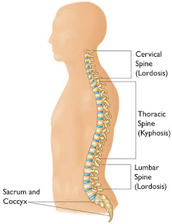
In the belly and for a time frame following birth, an infant's spine is formed like the letter C. This bend is named an essential bend, which is Kyphotic.

During the time the child is figuring out how to lift his head and in the long run walk, muscles create. As strong quality and capacity is picked up, the infant's action will move body weight to the spine. Steadily optional bends create in the cervical and lumbar districts; Lordotic bends. These bends will keep on creating until developing stops.

Spinal bends are either kyphotic or lordotic. In an ordinary spine there are four sorts of spinal shapes critical to adjust, adaptability, and stress ingestion and appropriation.


Posture : Normal And Kyphosis 


It tends to be ordered in five classifications:

1. Scheuermann's Disease:

The adolescent type of hyperkyphosis. Characterized as an imperfection of the vertebral body development plate at the cortical dimension, brings about debilitating the vertebral body and causing wedging. It stops toward the finish of development, when the development ligament is never again dynamic.

2. Postural Kyphosis:

Most regular type of kyphosis. It is an aftereffect of poor stance and debilitated muscles and tendons of the spine without any vertebrae deformations obvious.

3. Inherent Kyphosis:

Bone deformity recognized during childbirth. Kyphosis edge will increment if not treated in time.

4. Age-related Hyperkyphosis:

Kyphosis point increments with age, bringing about age-related hyperkyphosis. In spite of the way that expanded kyphosis is one of the average aftereffects of osteoporosis, up to 70% of patients with age-related hyperkyphosis don't experience the ill effects of diminished bone mineral thickness. [2] The precise commonness of patients for non-osteoporotic hyperkyphosis is obscure, yet it is assessed that 10% to 45% of the general population matured more than 50 years are appropriately analyzed. Genderwise, thoracic hyperkyphosis increments to half in men and 65% in ladies beyond 65 years old years.

5. Neurological Hyperkyphosis:

Can be an aftereffect of the loss of motion of muscular strength.

CAUSES:-

Kyphosis happens when the vertebrae in the upper back become more wedge molded.

Irregular vertebrae can be brought about by:

Cracks.

Broken or squashed vertebrae (pressure cracks) can result in ebb and flow of the spine. Mellow pressure breaks regularly don't deliver recognizable signs or indications.

Osteoporosis.


This bone-diminishing issue can cause spinal bend, particularly whenever debilitated vertebrae result in pressure cracks. Osteoporosis is most regular in more seasoned ladies and individuals who have taken corticosteroids for significant lots of time.

Circle degeneration.

Delicate, round circles go about as pads between spinal vertebrae. With age, these circles dry out and recoil, which regularly intensifies kyphosis.

Scheuermann's infection.

Additionally called Scheuermann's kyphosis, this malady normally starts during the development spurt that happens before pubescence. Young men are influenced more regularly than young ladies.

Birth deserts.

Spinal bones that don't grow appropriately before birth can cause kyphosis.

Disorders.

Kyphosis in kids can likewise be related with specific disorders, for example, Ehlers-Danlos disorder and Marfan disorder.

Malignant growth and disease medicines.

Malignant growth in the spine can debilitate vertebrae and make them increasingly inclined to pressure cracks, as can chemotherapy and radiation disease medicines.

Indications:-

On the off chance that you have kyphosis, the most straightforward to-perceive indication is an adjusted back. The outrageous bend in your upper spine will make you hunch forward.

Notwithstanding having an adjusted back, you may have these different side effects:

Gentle to extreme back torment

Back agony with development

Weariness

Delicacy and solidness in the spine

Forward stance of the head

Chest agony or trouble breathing (serious cases)

Contrast in shoulder tallness

Tight hamstrings (muscles in the back of your thighs)

Conclusion:-

X-Ray or CT Scan.

X-beams can decide the level of ebb and flow and distinguish deformations of the vertebrae. A CT output may be suggested if your specialist needs increasingly point by point pictures.

X-ray.

These pictures can distinguish contamination or a tumor in your spine.

Nerve tests.

In the event that you are encountering deadness or muscle shortcoming, your specialist may prescribe tests to decide how well nerve driving forces are going between your spinal line and your limits.

Bone thickness tests.

Low-thickness bone can compound kyphosis.

EXAMINATION :-

Physical Exam:

During the physical test, the specialist takes a gander at your back and feels your spine. The person will feel for variations from the norm. Additionally, this piece of the physical test—called palpation—can uncover delicate spinal muscles or delicate hamstring muscles. The specialist will push on your muscles to check whether there is any delicacy.

Kyphosis is best observed from the side, both standing straight and bowing forward.

The specialist may complete an Adam's Forward Bending Test: in that, you curve forward at the midsection. The specialist will search for an adjusted bend (progressively characteristic of postural kyphosis) or an increasingly rakish bend. The precise bend can be known as a gibbus distortion, and it's simpler to see when you curve forward. It's additionally simpler for the specialist to see thoracolumbar kyphosis when you twist forward (thoracolumbar kyphosis is a strange bend in the area where the thoracic and lumbar spines meet).

To test your scope of movement, your specialist will request that you curve forward, in reverse, sideways, and to wind. A few people with kyphosis think that its difficult to do these developments.

kyphosis cobb's edge:

Sensory system Exam:

Next, your specialist will test your sensory system. The sensory system incorporates the mind, spinal string, and nerves that broaden like tree limbs through the body. The nerves convey messages to and from the cerebrum to control your body. The specialist will inquire as to whether any piece of your body is difficult, shivering, numb, or frail. Since nerves make everything work in the body, the specialist will likewise inquire as to whether you can go to the washroom regularly.

Pneumonic capacity tests.

On the off chance that the bend is extreme, your specialist may arrange aspiratory capacity tests. These tests will help decide whether breathing is confined in light of lessened chest space.

Avoidance:-

Advancement of postural kyphosis can be anticipated with keeping up perfect stance, fortifying both stomach and back muscles, and keeping up perfect body weight.

TREATMENT:-


1.MEDICAL :

Torment relievers.

On the off chance that over-the-counter drugs —, for example, acetaminophen (Tylenol, others), ibuprofen (Advil, Motrin IB, others) or naproxen sodium (Aleve) — aren't sufficient, more grounded agony meds are accessible by remedy.

Osteoporosis meds.

Bone-fortifying meds may help counteract extra spinal cracks that would decline your kyphosis.

2.SURGICAL:


SPINAL FUSION:

Specialists perform spinal combination as a feature of each spinal reconstructive medical procedure in individuals with grown-up scoliosis or kyphosis. Spinal combination balances out the spine by forever consolidating two vertebrae, wiping out development between them. Normally, little bits of bone taken from different pieces of the body, called "joins," are set in the middle of vertebrae to enable them to combine. In time, new bone becomes over the unite. The principle occupation of the screws and poles set during medical procedure is to hold the vertebrae emphatically together while the bone development happens.

Spinal combination might be performed to join at least two vertebrae together. Extreme scoliosis or kyphosis may require combination of different vertebrae so as to balance out and fix the spine.

3.BRACING:

Propping is possibly suggested when the hyperkyphosis is never again reversible through activities since it is excessively hardened or in light of the fact that activities have officially demonstrated inadequate. It's best as a physiotherapist to decide for exercise treatment, since it's less intrusive and is along these lines a superior first line choice.

Propping is a significant component in the physical treatment of thoracic hyperkyphosis, yet it gives just helpful results related to active recuperation. The blend of both, decreases the spinal firmness. Latent supporting without the expansion of non-intrusive treatment, doesn't have any impact on the thoracic spine.
A couple of kinds of supporting for thoracic hyperkyphosis are referenced beneath:

A Milwaukee Brace is the most widely recognized prop utilized by patients with a thoracic hyperkyphosis. This support has back cushions pushing anteriorly on the kyphosis. The neck and the pelvis are constrained by different fragments of the support. The patients should wear this prop 23 hour daily for 1-2 years.

The consequences of the Lyon Antikyphosis Brace were palatable in many patients with thoracic hyperkyphosis.


Spine And Posture
Spine And Posture


Likewise another supporting structure called the Kyphologic Brace has been appeared to have a decent in-prop adjustment at normal.

Another sort of support utilizing the two 3-point weights.


Measurement Kyphosis

Thursday, February 28, 2019

Home Visit Physiotherapy Treatment : Odhav Ahmedabad

Physiotherapy Treatment At Home Visit Service in Ahmedabad 

Physiotherapy Treatment At Home in Odhav And Nearby Area Of Ahmedabad, is Required Mainly Bed Ridden Patient Which Are Unable to Move And Sit, Stand , Walk Or Required Assistance. This Kind Of Patient Totally Dependent On Family And Daily Require Physiotherapy Exercise For Complete Rehabilitation Of Patient . Experienced Physiotherapist First Assess The Patient And Explained Relative About Patient Physical Condition, Treatment Procedure And Relative Outcome Of Treatment And Exercise.

Rehabilitation And Physiotherapy Treatment


Home Visit Treatment is More Useful in This Case Mostly This Kind Patient Are Paralytic Condition Like Hemiplegia, Paraplegia, Quadruplegia, Motor Neurone Disease, GBS , Other Disease That Are Related. Extra Care, Motivation , Proper Nursing Staff Also Require in Some Patient.

Home Visit Physiotherapy Odhav


Physiotherapy Center Near Odhav Area is Samarpan Physiotherapy Fitness And Rehabilitation Clinic Located in Four Places in East Ahmedabad , From Takshila Road To Nirant Char Rasta Vastral Branch And Other is At Rabari Colony Char Rasta, Amaraiwadi is Also Near To Odhav From Rajendrapark Char Rasta, One Branch Near India Colony Road Bapunagar.

Physiotherapy Appointment Home Care Treatment 


Special Community Based Rehabilitation Programme From Physiotherapist , Nursing Staff, Occupational Therapist is Useful Mainly And Sometime Speech Therapy Also.
Mobile Physiotherapy Clinic Near Tollnaka Bapunagar is Also Near To Odhav. Mobile Physiotherapy Clinic is Specially Useful in Home Visit Treatment.

Physiotherapy clinic in Vastral Near Nirant Char Rasta :

Samarpan Physiotherapy clinic Vastral Branch
                         
B-04, Shivalik Bunglow, Near. Metro Pillar No. 156
Near. Ratanpura Gam,
Madhav School Road                
Vastral Road 
Ahmedabad

Time :    

Morning : 8: 30am to  12pm
Evening  : 4 :00pm  to  8 :00pm.

Dr.  Nitesh Patel ( BPT, MIAP)
M:  9898607803
   :  7777976968

Bapunagar clinic address : 
  
Samarpan Physiotherapy clinic
Surjit Society,Behind. Nilkanth Dairy,
Nr.Ramaji Mandir, Near. Satsangi school
India Colony Road, Bapunagar, Ahmedabad.

Clinic Time :    

Morning : 8 : 30 am to  12: 00pm
Evening : 4 : 00pm  to  8 : 00pm.
 
                          DR.  NITESH PATEL ( BPT, MIAP)
                                     M:  9898607803
                                         :   9724757508

                          
Amaraiwadi area Near. Rabari Colony Char Rasta

Samarpan Physiotherapy Clinic
Bansidhar Society,Opp.Revabhai Shopping Centre,
B/H.Bansidhar Medical Store,
Vastral Road, Amaraiwadi, Ahmedabad.

Clinic Timing  :

Morning : 8 :00am to  12 :00pm
Evening : 4 :00pm  to  8 :00pm.
 
Dr. Nitesh Patel ( BPT, MIAP) M:  9898607803
Dr.  Komal Chauhan - Physiotherapist
                              Mo No. : 6352845017

India Colony Road : 

Mobile Physiotherapy Clinic.
B-01, Jagatnagar Society, Opp.Shaktidhara Society.
India Colony Road to Tollnaka,
Bapunagar, Ahmedabad.

TIME:   Morning Session : 9:00am to  12:00pm  ,  
Evening Session :  4:00pm  to  8:00pm.

Nikol Naroda Road :

Samarpan Physiotherapy Clinic
11, Vedant Bunglow, Opp.Radhe 2 Bunglow,
Near.Fortune Circle,
Near. Haridarshan Char Rasta
Behind. Shalby Hospital
Nikol New Naroda Road
New India Colony Road
Ahmedabad.

Timing : 

Morning :  9 :00 am to 1 :00 pm
Evening  :  4 :00 pm to 8:00 pm

Dr. Nitesh Patel : Physiotherapist.

Mo no : 09898607803

Dr. Varsha Patel - Physiotherapist.
Dr. Dharti Patel - Physiotherapist
Dr. Shivangi Chauhan - Physiotherapist.